Brain Crash Course – Its Structure and What Meditation Changes

 · 10 min read
While browsing through old blog articles, I noticed something: parts of the brain keep getting mentioned. It immediately took me back to my first semester at university: amygdala here, hippocampus there, prefrontal cortex everywhere. Until I ironically started to wonder whether the hippocampus was supposed to be a hippo steak and the amygdala a new mosquito repellent.
That’s why today I’m giving you a little crash course: together we’ll travel from the oldest parts of your brain all the way up to the cerebrum. That way you’ll have read it all once and can look things up later whenever you need to. And so that I don’t lose you along this long journey, at the end I’ll also reveal which regions have been shown to change through meditation and mindfulness. Let’s get started straight away with our first monster of a word: “rhombencephalon”.
Rhombencephalon (Hindbrain) – The Ancient Foundation
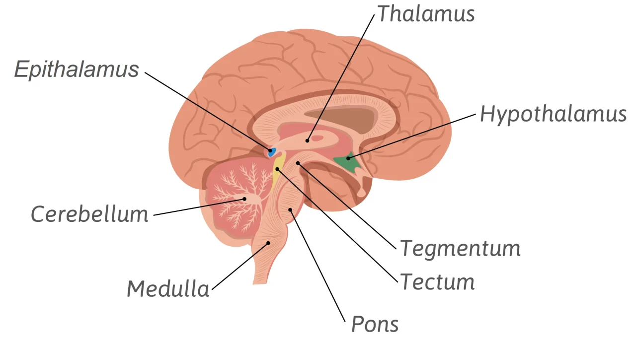
The hindbrain is the lowest and, in evolutionary terms, the oldest part of your brain. This is where the basic life functions are located: heartbeat, breathing, reflexes and the sleep-wake cycle. Almost all the nerve pathways between body and brain run through this area, which is why it has such a big influence. When it is more active, you feel more awake and alert. When it is less active, you become sleepy. The hindbrain consists of two large sections:
- Myelencephalon (Medulla Oblongata) – The Gateway
This is where your spinal cord ends and your brain proper begins. However, the myelencephalon also has tasks of its own: it regulates heartbeat, blood pressure and breathing, and controls reflexes such as swallowing or coughing. All of this works even when you are unconscious - Metencephalon – The First Building Block
For one thing, the metencephalon includes the cerebellum – an ancient structure found in all vertebrates and built like a tiny mini-brain. It coordinates movements, both gross and fine motor, and regulates muscle tone. It also plays an important role in learning through conditioning.
The second important structure of the metencephalon is the pons, also known as the bridge. The name says it all, as it connects your hindbrain with the midbrain and the cerebrum. At the same time, it has tasks of its own: it controls balance, REM sleep and a large part of the facial muscles.
Mesencephalon (Midbrain) – The Bouncer
The midbrain sits right between the old hindbrain and the large, modern forebrain. In fish and reptiles it is still the central control centre for movement; in humans, higher brain regions have taken over many of its tasks. Despite its small size, the midbrain remains indispensable. Damage to this region would have serious consequences, because it contains fundamental relay stations for movement, attention and sensory processing.
It can be further divided into the tectum and the tegmentum.
- Tectum – For Reflexes
In the tectum, motor activity is linked with sensory impressions. This allows you to react in a flash, for example by tracking a moving object with your eyes, repositioning your hands to grab something, or turning your head towards a sound. That’s because initial auditory information is processed here too – the tectum, for example, calculates the location of a sound source based on the differences in timing and volume between your two ears. - Tegmentum – A Dopamine Factory
Running through the tegmentum is the substantia nigra, a small but crucial cluster of nerve cells that produces large amounts of dopamine. If this structure is destroyed – as in Parkinson’s disease – it leads to tremors, movement disorders and often depressive symptoms as well.
Prosencephalon (Forebrain) – From Animal to Human
With the prosencephalon, we arrive at the largest and most highly developed part of the brain. It makes up around 80% of your total brain mass and is responsible for abilities that clearly set you apart from the rest of the animal kingdom. In evolutionary terms, this area is comparatively young. The prosencephalon is divided into two large areas: the diencephalon and the telencephalon.
Diencephalon (Interbrain) – Your Operating System
The diencephalon is a central hub of your brain. This is where vital bodily functions are regulated and coordinated so that your organism stays stable and runs smoothly.
- Thalamus – Your Gateway to Consciousness
The thalamus is the sensory control centre of your brain. Every second, millions of sensory impressions rain down on you. So that you don’t get overwhelmed by them, your thalamus filters at lightning speed which information gets passed on to your cerebrum and which is screened out. That’s why it is often called the “gateway to consciousness” – whatever gets through here reaches your conscious awareness; whatever it discards goes unnoticed. - Hypothalamus – Your Inner Thermostat
The hypothalamus is the endocrine control centre of your brain. It produces hormones itself and at the same time controls hormone production in other organs. Although it is barely bigger than an almond, it influences central vital and metabolic functions: temperature regulation, hunger, thirst, sexual function, growth, stress responses, mood and much more. - Epithalamus – A Hand on the Brake
The epithalamus includes, among other things, the pineal gland, which controls your sleep-wake cycle through its melatonin production. This area also contains other structures whose functions are not yet fully understood. However, much suggests that they are involved in correcting behaviour: in response to negative feedback, they reduce dopamine production in order to prevent or weaken harmful behaviour.
Telencephalon (Cerebrum) – Instinct Becomes Consciousness
The telencephalon is the most complex part of your brain. This is where language, thought, emotions, memory and everything that makes up your conscious experience arise. It can be divided into several large functional areas.
- Basal Ganglia – Your Autopilot
Among other things, the basal ganglia are the seat of your procedural memory. This is where movement sequences and routines are stored, so you don’t have to relearn them every time. Walking, cycling, playing the piano or even opening a plastic bottle run automatically once you’ve practised them – without much mental effort. - Amygdala – Inside Out
Your amygdala holds many innate emotional response patterns, especially those linked to fear, anxiety, aggression and sexual behaviour. It controls physical reactions such as heart rate, breathing rate, freezing or the release of stress hormones, and also influences behaviour patterns such as screaming or certain facial expressions. However, it is not solely responsible for your emotions – they are far too complex for that. Emotions arise from the interplay of many brain regions that communicate with one another via the limbic system. - Hippocampus – The Source of Your Memories
The hippocampus is central to declarative memory – that is, everything you can consciously remember and put into words. During the day, it records experiences with great accuracy. At night, it stores this information in the cerebral cortex and coordinates where which content is filed. If your hippocampus stopped working, you would live in the past forever, with everything newly experienced – forgotten. - Cerebral Cortex – The Part Reading This Sentence Right Now
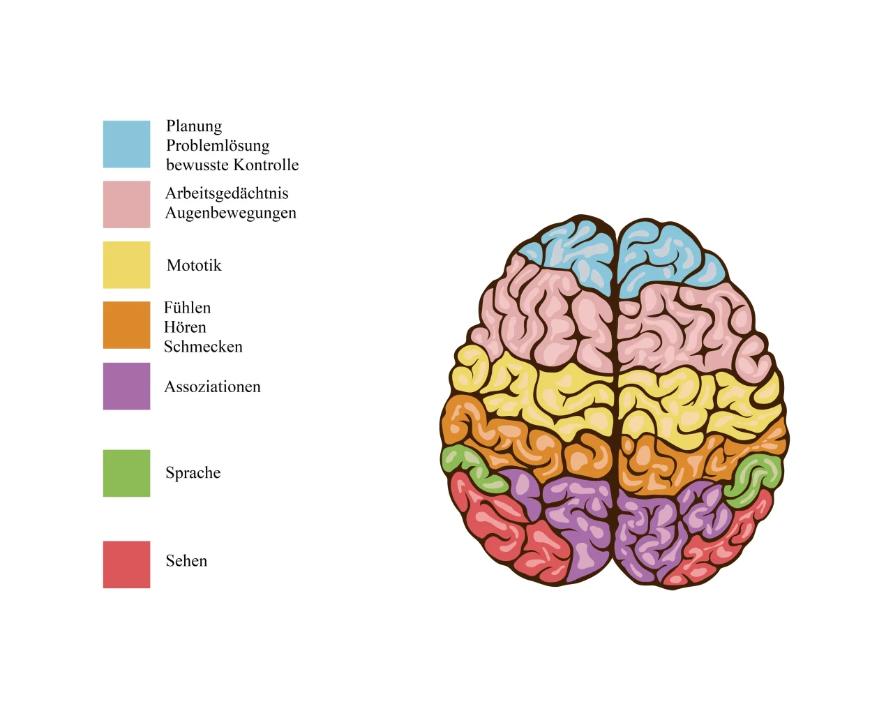
The folded grey surface you’ve surely seen many times in pictures of the brain is the cerebral cortex. Almost all higher cognitive functions arise here. It also holds a large part of your long-term memory, especially in the form of semantic information. Because of its complexity, the cortex is further divided into different areas. Greatly simplified:
- Prefrontal cortex
Responsible for planning, working memory, problem-solving, decision-making and conscious control. - Motor cortex
Enables you to consciously control your movements and muscles. - Somatosensory, visual, auditory, olfactory and gustatory cortex
These areas process the sensory impressions that your thalamus hasn’t filtered out. - Broca’s and Wernicke’s areas
They help you understand (Wernicke) and produce (Broca) language. - Association cortex (parietal)
Links information together and enables abilities such as personality, facial recognition, spatial thinking and vocabulary.
Glial Cells and Nerve Cells – Workers and Thinkers
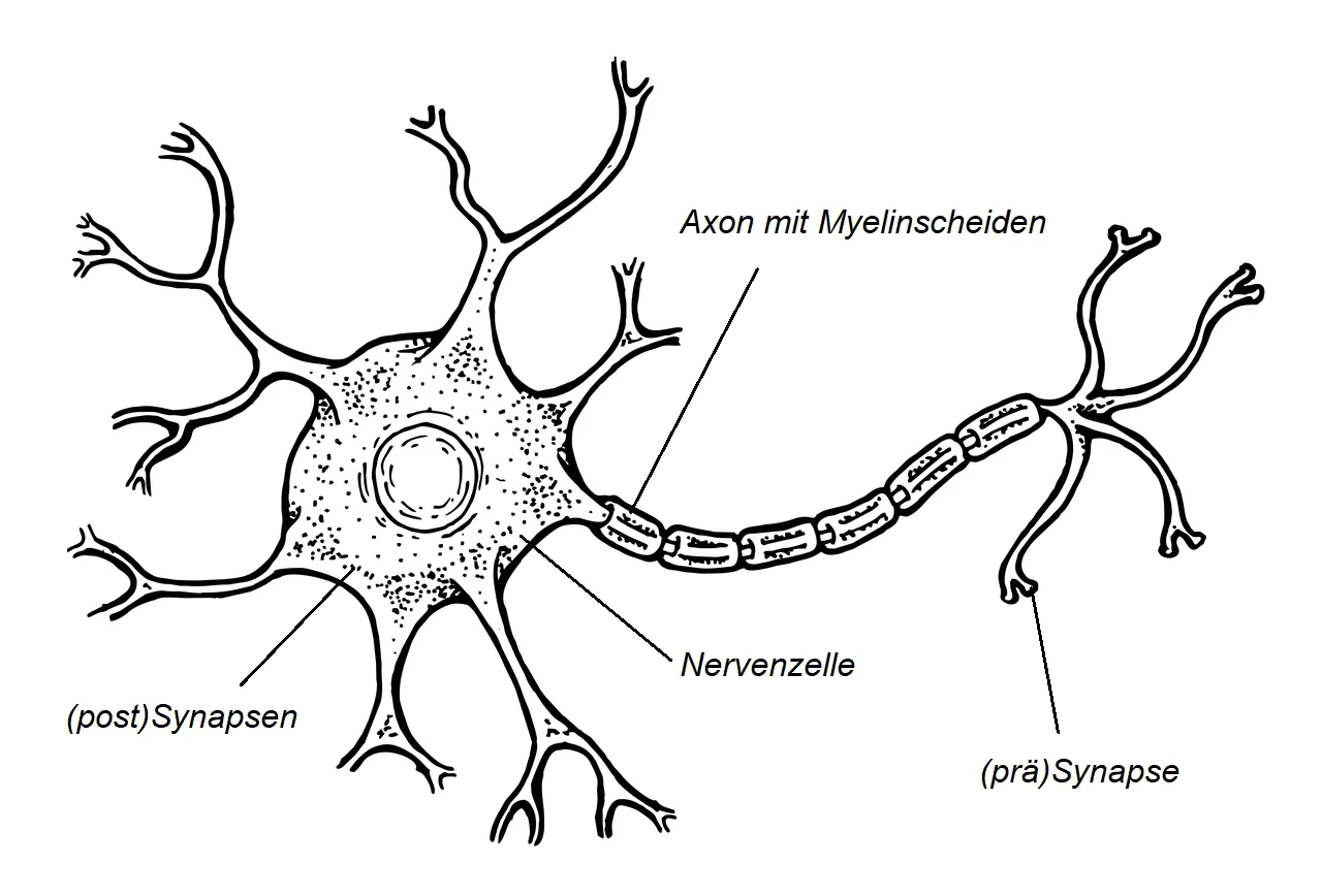
For a long time, glial cells were thought to be nothing more than “glue” between the neurons. Yet they make up about half of your brain cells. They perform a kind of maintenance and supply role without which your nervous system could not survive. Their tasks range from repairing minor injuries and regulating the blood-brain barrier to supplying neurons with oxygen and nutrients. They also dispose of dead cells and waste products, stabilise the tissue and form the myelin sheaths that wrap around certain axons and greatly speed up their signal transmission.
The other roughly 50 per cent of your brain cells are nerve cells (neurons). They are highly specialised, long-lived and have striking, long extensions – the axons. Through these, they connect with other nerve cells. At the points of contact, the presynapses, electrical impulses are translated into chemical signals and passed on, usually via neurotransmitters such as dopamine, serotonin or glutamate. At the postsynapses, these then trigger an electrical signal again, the action potential. A single neuron can have up to 10,000 connections to other neurons. Together they form the gigantic network in which this very sentence is taking shape in your head right now.
Grey and White Matter – Country Road vs. Motorway
Grey matter consists of large clusters of nerve cell bodies. It is called grey matter because, when fresh and viewed with the naked eye, it looks like a greyish-brown gel.
White matter consists of axons wrapped in myelin sheaths. These myelin sheaths act like insulation and ensure that electrical signals are transmitted much faster and more reliably – like on a motorway.
However, myelinated axons have their downsides:
They take up a lot of space and consume a lot of energy. That’s why your brain uses myelin selectively, for particularly important information or for signals that need to travel over longer distances.
How Does Thinking Work? – When Neurons Dance
The principle of thinking is best explained with a simple example: perception.
Imagine you’re a little baby and the world is still completely new. There’s an object in front of you. You reach for it, look at it, press it, put it in your mouth: green, hard, angular, tastes of wood. Mummy says: “green building block” – whatever that means…
All these sensory impressions travel to the brain and activate a particular group of neurons there. These nerve cells start exchanging electrical signals and strengthen their connections. The next time you see something green, hard and angular, exactly the same neurons fire again. You recognise the pattern – now all you’d need to do is be able to say “green building block”. How did Mummy do that again?
Then something new happens: you find another building block. Again hard, angular, tastes of wood – but this time not green. The existing pattern doesn’t fit. The “green” neurons stay quiet, other neurons kick in. After a short while, you grasp the principle: there are red, blue, yellow and green blocks. Each colour creates a slightly different activity pattern in the brain. So it quickly becomes clear: information never sits in a single nerve cell. It arises from the interplay of many neurons that together perform a very specific “dance of electrical signals”.
And exactly this principle applies to everything – whether a building block, an emotion, a daydream or a fractional Chern insulator with subsystem-protected type II fracton orders. Everything is an individual activity pattern made up of millions of neurons firing simultaneously and communicating with each other. This activity can be measured with an EEG. Depending on its strength and spread, it shows up there as alpha, beta or gamma waves.
What Do Meditation and Mindfulness Change in the Brain?
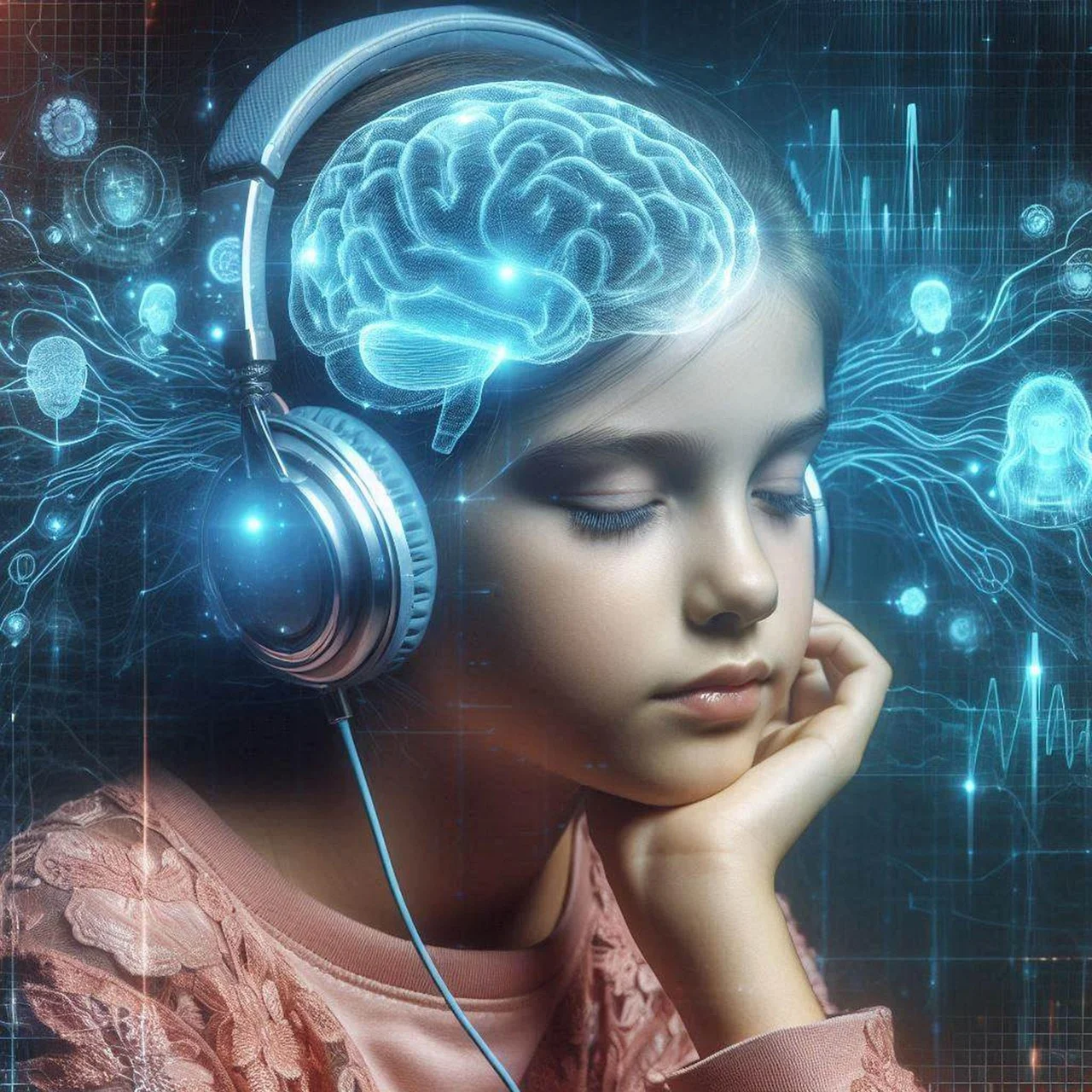
Meditation affects your brain much like strength training affects your body. In several brain regions, the amount of your grey and white matter increases, especially in the cerebral cortex. As a result, there are not only more nerve cells at work, but they also work together more efficiently. At the same time, the age-related loss of brain tissue slows down – you stay mentally fit for longer. The areas that benefit most are those responsible for attention, memory, sensory processing and emotion regulation.
Another striking finding: the overall activity of your brain decreases. At first that sounds as if you’d become sleepy or sluggish – but in fact the opposite happens. Meditation leads to greater clarity, alertness and mental sharpness. The reason: your brain produces more high-frequency gamma waves (30-100 Hz), which are associated with intense perception and high cognitive performance. So it doesn’t become less active; it simply uses its resources far more efficiently, precisely and in a more focused way.
Mindfulness training also changes the structure of your amygdala – the region that responds to fear and stress. Regular meditation causes certain parts of the amygdala to shrink, which leads to fewer negative feelings. Other areas, on the other hand, can grow, for example through metta meditation. This improves your ability to perceive other people’s emotions, to feel compassion and to act empathetically.
Your hormone balance also shifts in a positive direction. Dopamine and melatonin rise, while cortisol and noradrenaline fall. The result: more calm, a better mood and an overall more stable emotional balance. The increase in grey matter in the hippocampus further strengthens this effect. It promotes a calmer, more self-assured self-image that is more firmly anchored in the here and now.
Neurotransmitters
Phew – that’s it for now. Hats off to you if you’re still here after this long and rather dry lesson. If you’re still hungry for more, here’s an article that takes a closer look at the neurotransmitters mentioned. But first, I’m off to make myself a cup of tea…
Tom L. (psychology student)
Deepen your meditation practice with Mindfulife
Meditation can be more than a short break in your everyday life. If you would like to understand more deeply how meditation works, how to build a steady practice of your own and how to guide others mindfully, our meditation teacher training is the next step. There you will not only get to know meditation in theory, but also develop more confidence, clarity and trust in your own practice.
Would you like to find out more about the training first? In our weekly webinar you will get an insight and learn more about what your path to becoming a meditation teacher could look like.
For regular support in your everyday life, you can also use our online meditation studio and join guided meditations flexibly via Eversports.
We look forward to accompanying you on your journey.
You can find more mindfulness inspiration on our channels too
References
Damiani, Daniel; Nascimento, Anna; Pereira, Letícia . (2017). Funções corticais cerebrais – o legado de Brodmann no século XXI. Arquivos Brasileiros de Neurocirurgia: Brazilian Neurosurgery, (), s-0036-1597573–. doi:10.1055/s-0036-1597573
Esch, T. (2014). Die neuronale Basis von Meditation und Achtsamkeit. Sucht, 60(1), 21–28. doi:10.1024/0939-5911.a000288
Güntürkün, O. (2018). Biologische Psychologie (2nd edn) [EPUB]. doi:10.1026/02941-000
Larry W Swanson. (2000). What is the brain?, 23(11), 0–527. doi:10.1016/s0166-2236(00)01639-8
Michaelsen, M.M., & Esch, T. (2021). Die neuronale Basis von Meditation und Achtsamkeit im Bildungskontext. In Iwers, T. & Roloff, C. (Hrsg.), Achtsamkeit in Bildungsprozessen (S. 61-75). Springer. https://doi.org/10.1007/978-3-658-30832-2_5
Rossbach, G. (2019). Aus der Hirnforschung: ein Loblied auf Meditation. In: Glücksorgan Gehirn. Springer, Berlin, Heidelberg. https://doi.org/10.1007/978-3-662-57729-5_17
Rigos, A. (2025, March 27). Evolution des gehirns. geo.de. https://www.geo.de/natur/tierwelt/das-gehirn-evolution-des-gehirns-30192468.html